Oxygen-carrying reserve
Plasma volume expands more than red-cell mass, producing physiologic dilutional anemia. Interpret against gestational age, symptoms, bleeding, and prior values.
After completing this chapter, you should be able to interpret pregnancy-specific laboratory trends, recognize high-risk patterns, identify preanalytic limitations, use focused diagnostic testing within scope, and communicate how results change the transport plan.

Question
What clinical decision will this test inform?
Pregnancy baseline
Is the expected normal range different?
Trend
Is the value rising, falling, or stable?
Timing
Was it drawn before or after treatment or bleeding?
Action
Does it change treatment, destination, or urgency?
A 31-year-old patient at 35 weeks is transferred for severe hypertension and right-upper-quadrant pain. Platelets have fallen from 168,000 to 92,000/µL in six hours, creatinine has risen from 0.6 to 1.2 mg/dL, AST is more than twice normal, and the fetal tracing is worsening. No single number tells the whole story. The trend shows evolving multisystem disease and a narrowing margin for safe transport.
Plasma volume expands more than red-cell mass, producing physiologic dilutional anemia. Interpret against gestational age, symptoms, bleeding, and prior values.
Pregnancy and labor can raise the white count. Leukocytosis alone does not diagnose infection; fever, source, differential, lactate, perfusion, and trend matter.
Mild gestational thrombocytopenia is common, but a rapid fall or count below 100,000/µL is concerning in hypertensive disease and hemorrhage.
| Test | Concerning pattern | Clinical meaning |
|---|---|---|
| Platelets | Falling trend; below 100,000/µL | Severe feature, consumption, bleeding and neuraxial implications |
| Creatinine | Above expected pregnancy baseline; >1.1 mg/dL or doubled baseline | Renal involvement and altered medication clearance |
| AST/ALT | Elevation, especially about twice normal with symptoms | Hepatic involvement; consider HELLP and other liver disease |
| Urine protein | 24-hour ≥300 mg or spot P:C ≥0.3 | Supports proteinuria; absence does not exclude severe features |
| LDH, bilirubin, haptoglobin, smear | Pattern consistent with hemolysis | Supports HELLP or another hemolytic process |
Uric acid may reflect disease severity in some contexts but is not a stand-alone diagnostic test. The transport clinician should report the pattern: symptoms, blood pressure, platelets, renal function, liver tests, urine output, and fetal trajectory.
Yes. Severe features can establish high-risk disease even without proteinuria.
Helpful when factor depletion, anticoagulant effect, liver dysfunction, or disseminated intravascular coagulation is suspected. Normal results do not exclude early coagulopathy.
Normally elevated in pregnancy. A value near the nonpregnant normal range can be relatively low during active hemorrhage; falling values are concerning.
Where available, TEG or ROTEM may provide rapid functional information and support targeted blood-product therapy.
Confirm type, antibody screen, crossmatch status, products available, transport cooler requirements, and massive-transfusion pathway.
Pregnancy can develop diabetic ketoacidosis at lower glucose concentrations than many clinicians expect. Do not exclude DKA because the glucose is only moderately elevated. Normal pregnancy also lowers PaCO₂ through increased minute ventilation; a PaCO₂ near 40 mm Hg may represent relative hypoventilation.
Confirm current type and whether the patient is Rh-negative.
Identify clinically significant red-cell antibodies that affect crossmatching and fetal anemia risk.
ABO/Rh plus antibody screen; useful for patients who may need blood.
Tests compatibility with selected donor units and supports rapid issue.
The Kleihauer-Betke test estimates fetal red cells in maternal circulation and can help quantify fetomaternal hemorrhage and guide Rh immune globulin dosing. It has sensitivity and interpretation limitations and must not replace fetal monitoring, obstetric evaluation, or clinical judgment. A negative test does not exclude placental abruption or all fetal-maternal bleeding.
CDC recommends GBS screening during the 36th or 37th week of each pregnancy. During transport, verify the current pregnancy result, allergy history, intrapartum antibiotics already given, dose times, rupture duration, gestational age, and maternal temperature. GBS status is one part of neonatal-risk assessment, not a substitute for evaluating chorioamnionitis or sepsis.
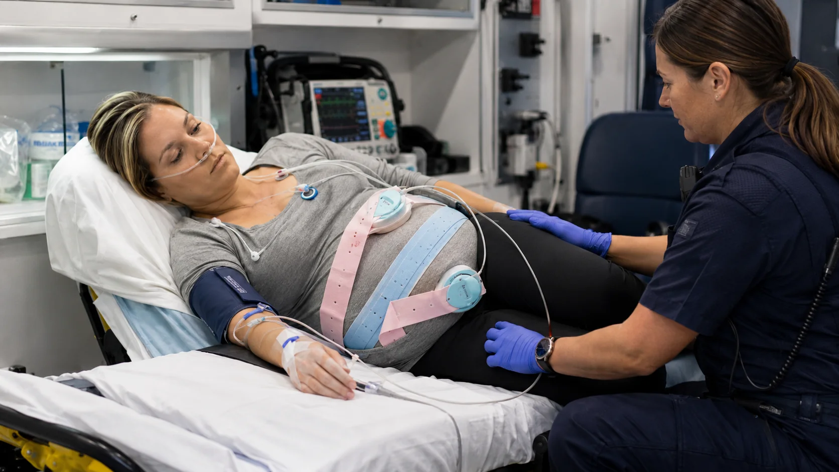
Fetal cardiac activity, presentation, gross fluid assessment, placental location, and selected biometry when trained.
Gross ventricular function, pericardial effusion, right-heart strain pattern, and volume-response clues.
B-lines, pleural sliding, effusion, and focal consolidation patterns.
Free fluid and focused causes of hypotension, while recognizing pregnancy-related anatomy and test limits.
Use ECG for chest pain, syncope, dysrhythmia, severe electrolyte disturbance, medication toxicity, or cardiopulmonary symptoms. Clinically necessary radiography, CT, MRI, or contrast-enhanced studies should not be withheld solely because the patient is pregnant when maternal benefit outweighs risk. Stabilize first, use the most appropriate test, and communicate gestational age and clinical urgency.
Record collection time and whether it was before or after fluids, medication, seizure, hemorrhage, or delivery.
Identify arterial, venous, line draw, urine, swab, or point-of-care source.
Consider hemolysis, dilution from IV fluids, clotting, mislabeling, and transport delay.
Compare with prior results rather than reading one value in isolation.
Report what changed and how it alters treatment, urgency, or destination.
The patient has severe-range hypertension, RUQ pain, platelets 118,000/µL, creatinine 0.9 mg/dL, and AST mildly elevated. The crew records collection times and requests the prior trend.
Repeat results show platelets 92,000/µL, creatinine 1.2 mg/dL, and AST more than twice normal. Urine output has fallen. The pattern is more important than any one value.
The team confirms magnesium and antihypertensive therapy, reviews blood-bank readiness, increases reassessment frequency, and sends the updated results to the receiving center.
Worsening fetal status and multisystem maternal laboratory deterioration prompt immediate discussion of the nearest facility capable of emergency delivery, anesthesia, blood-bank support, and neonatal stabilization.
Immediate rationales are shown in study mode, and your score is stored locally.