Threats
Airway, breathing, circulation, hemorrhage, seizure, mental status, delivery.
After completing this chapter, you should be able to perform a structured maternal primary and secondary assessment, interpret pregnancy-specific trends, identify early warning findings, recognize shock and cardiopulmonary deterioration, integrate basic fetal status, and communicate time-linked changes during transport.
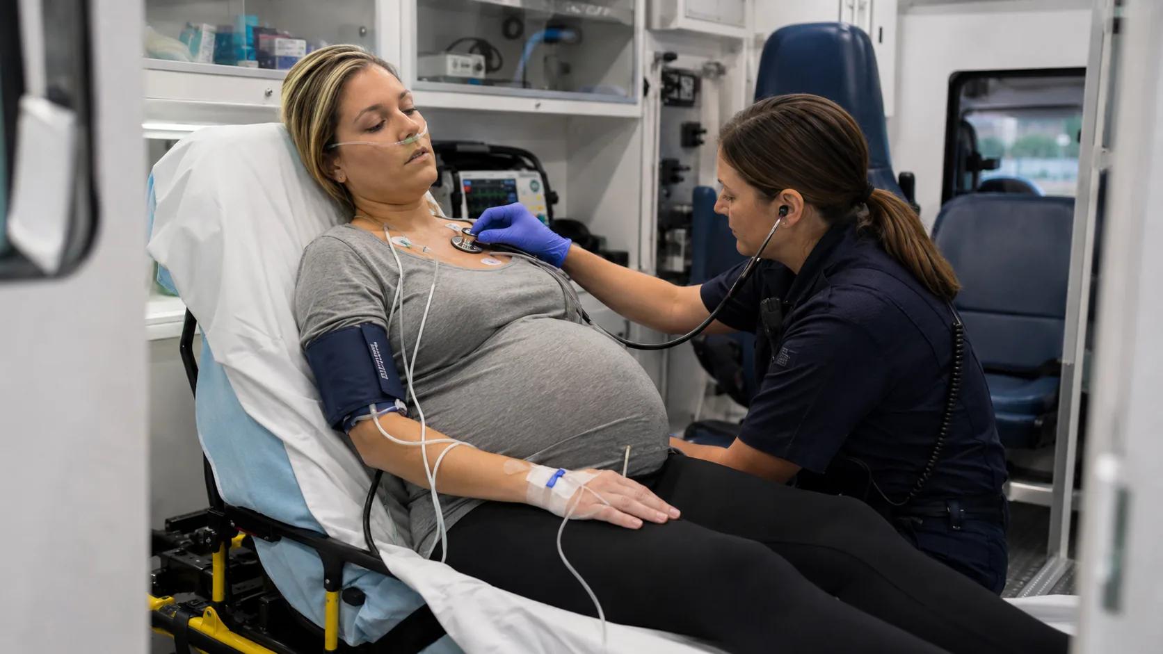
Is there an immediate threat?
Airway, oxygenation, hemorrhage, seizure, shock, or imminent birth.
Is position contributing?
Relieve aortocaval compression when appropriate.
What is changing?
Compare trends, symptoms, examination, and fetal response.
What mechanism fits?
Hemorrhagic, septic, cardiogenic, obstructive, or mechanical.
Does the plan still fit?
Continue, pause, divert, deliver, or add resources.
A 28-year-old patient at 34 2/7 weeks is being transferred for severe-range hypertension. Blood pressure is 166/112 mm Hg. She reports persistent headache and visual “sparkles.” Her face and hands are newly swollen, patellar reflexes are brisk, and ankle clonus is present. Twenty minutes into transport, respiratory rate rises to 30/min, SpO₂ falls to 92%, and bibasilar crackles develop. The physical assessment—not the referral label—shows worsening multisystem disease and possible pulmonary edema.
Airway, breathing, circulation, hemorrhage, seizure, mental status, delivery.
Relieve aortocaval compression and document the response.
Vital signs, perfusion, pain, urine output, respiratory and neurologic findings.
Uterine tone, contractions, bleeding, fluid, movement, basic fetal status.
Continue, pause, divert, prepare to deliver, or request resources.
History and examination occur simultaneously. A complete head-to-toe examination must never delay treatment of seizure, severe respiratory distress, major hemorrhage, shock, cord prolapse, or imminent delivery.
Distress, posture, behavior, pallor, diaphoresis, swelling, visible bleeding.
Ability to speak, respiratory pattern, accessory muscles, cough, stridor.
Orientation, agitation, lethargy, seizure activity, focal deficits.
Contraction behavior, urge to push, fluid, presenting part, fetal monitor.
Use the correct cuff, support the arm, and repeat unexpected results. Severe range is systolic ≥160 or diastolic ≥110 mm Hg.
A higher baseline may be physiologic, but persistent marked tachycardia needs an explanation.
A rising rate is often an early warning sign. Observe effort, speech, and trajectory.
New hypoxemia or oxygen requirement is abnormal and requires rapid evaluation.
38°C (100.4°F) or higher is an urgent warning sign and may affect fetal heart rate.
Low output may reflect hypoperfusion, renal injury, preeclampsia, or magnesium accumulation.
| Finding | Why it matters | Immediate response |
|---|---|---|
| SBP <90 or >160 | Shock or severe hypertension | Validate promptly; escalate and treat mechanism |
| DBP >110 | Severe hypertension | Immediate escalation per protocol |
| HR <50 or >120 | Dysrhythmia, shock, medication, infection | Reassess rhythm, perfusion, symptoms, and trend |
| RR <10 or >30 | Ventilatory failure or critical illness | Assess airway, oxygenation, ventilation, and cause |
| SpO₂ <95% | Maternal and fetal oxygen-delivery risk | Verify waveform, support oxygenation, evaluate cause |
| Confusion or unresponsiveness | Neurologic or systemic emergency | Immediate stabilization and differential diagnosis |
These are example maternal early-warning triggers used to prompt validation and escalation. They support rather than replace clinical judgment and local policy.
No. Compensated hemorrhage may be present before hypotension. Interpret skin, pulse pressure, mental status, bleeding, urine output, pain, and fetal trajectory.
Hyperreflexia and clonus may support neurologic irritability but do not independently diagnose preeclampsia. During magnesium therapy, decreasing or absent reflexes, respiratory depression, declining mental status, and oliguria raise concern for toxicity.
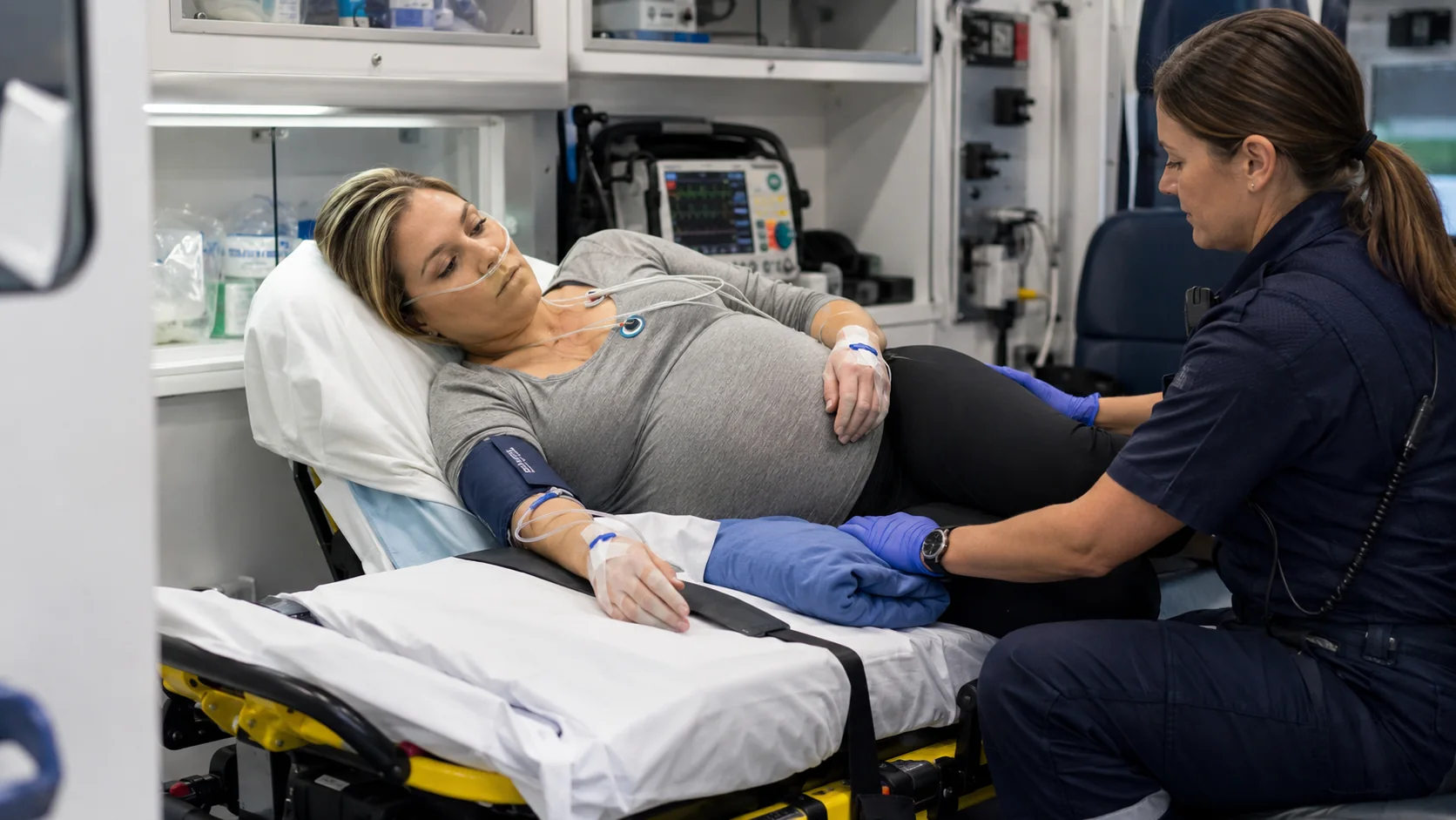
Crackles, hypoxemia, orthopnea, inability to speak normally, new oxygen requirement, or jugular venous distention are not explained by normal pregnancy alone. Sudden dyspnea with pleuritic pain, syncope, or unilateral leg symptoms requires consideration of pulmonary embolism.
Distention, scars, trauma, visible movement, bleeding, fluid, presenting part.
Location of pain, uterine tenderness, resting tone, contractions, rigidity.
Gestational age, fetal movement, fetal status, membrane status, prior surgery.
Do not perform routine digital vaginal examination in unexplained late-pregnancy bleeding before previa is excluded.
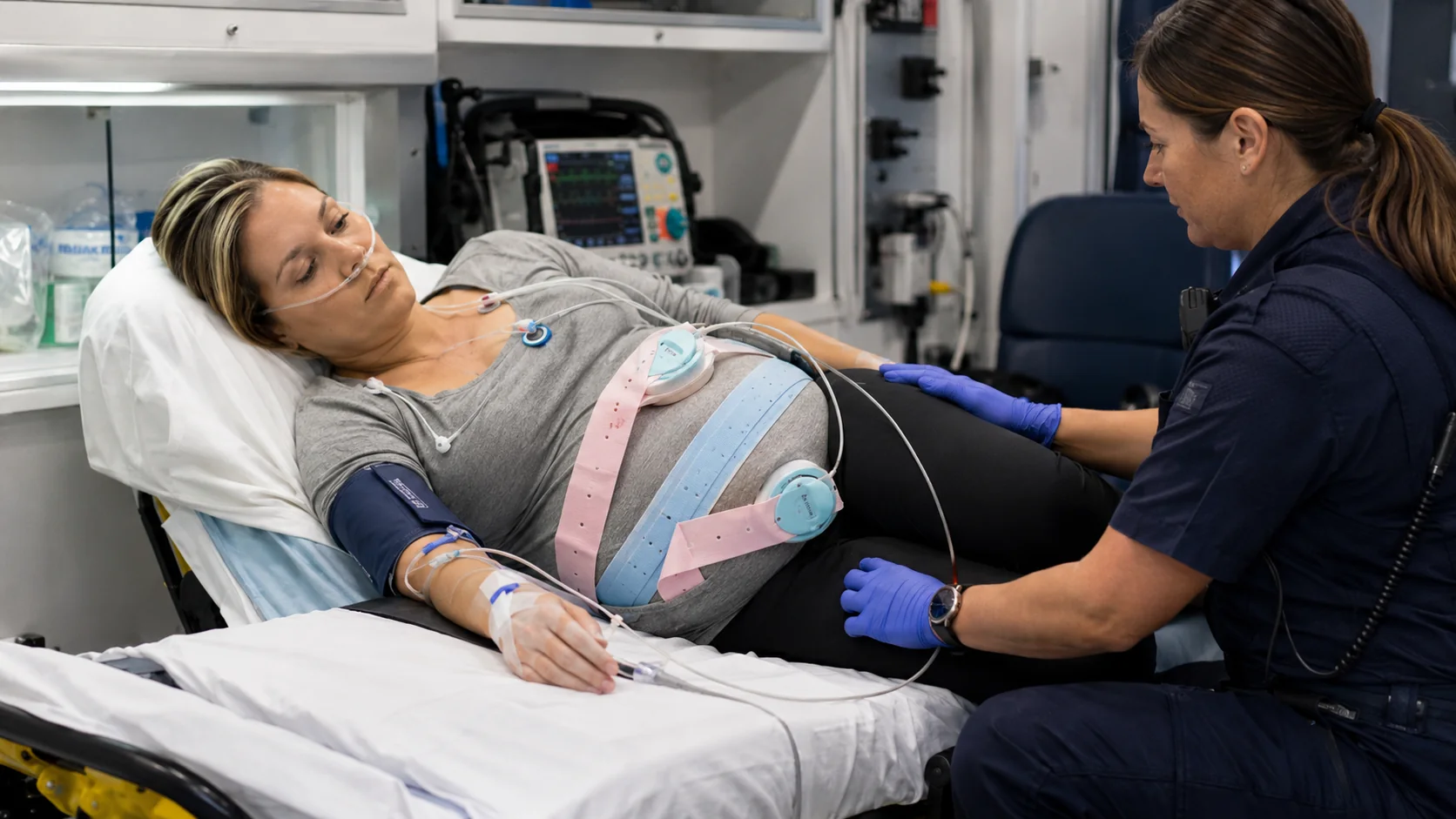
Painful bleeding, uterine tenderness, increased resting tone or rigidity, fetal deterioration; bleeding may be concealed.
Classically painless bright-red bleeding with a softer uterus; presentations vary and diagnosis requires imaging.
Sudden severe pain, shock, abnormal fetal status, loss of station, or concerning history of uterine surgery.
Onset, amount, progression, color, clots, pain, and concealed-bleeding clues.
Clear, blood-tinged, green/brown, foul-smelling, continuous, or intermittent.
Visible cord, presenting part, urge to push, rectal pressure, fetal deterioration.
Placental abruption, uterine rupture, retroperitoneal bleeding, or intra-abdominal hemorrhage may be concealed. Maternal perfusion and fetal status may worsen despite limited external blood.
Pallor, cool clammy skin, delayed refill, weak pulses, anxiety, confusion, narrowing pulse pressure.
Petechiae, bruising, or oozing from access sites may suggest thrombocytopenia or coagulopathy.
New unilateral swelling, warmth, redness, or tenderness is more concerning than symmetric dependent edema.
Persistent RUQ/epigastric pain, pleuritic chest pain, tearing back pain, flank pain with fever, or constant pain between contractions.
At minimum, integrate gestational age, fetal movement, fetal heart rate or recent tracing, contractions, membrane status, placental conditions, and the response to maternal stabilization. Detailed tracing interpretation is addressed in Chapter 10.
Tachycardia, pallor, cool skin, narrowing pulse pressure, bleeding or concealed loss, fetal deterioration.
Fever or hypothermia, tachycardia, tachypnea, altered mentation, suspected infection, abnormal perfusion.
Dyspnea, orthopnea, crackles, JVD, dysrhythmia, edema, chest symptoms, poor perfusion.
Sudden dyspnea, chest pain, syncope, hypoxemia, hypotension, unilateral leg findings, tension physiology.
Supine symptoms that improve with left tilt or uterine displacement.
After position change, loading, transfer, or major vibration.
After medication, infusion, oxygen, airway, or fluid adjustment.
After new pain, bleeding, fluid, contractions, dyspnea, or neurologic symptoms.
After a monitor or equipment alert—assess the patient, not only the device.
Time-link objective trends and notify the receiving system early.
The 34-week patient has severe-range hypertension, headache, visual symptoms, brisk reflexes, and clonus. The crew verifies the magnesium concentration and rate, confirms urine output, establishes seizure precautions, and reports the findings.
Respiratory rate rises to 30/min, SpO₂ falls to 92%, and bibasilar crackles develop. The team recognizes possible pulmonary edema rather than attributing dyspnea to anxiety or normal pregnancy.
Positioning and oxygenation are optimized, infusions and prior fluid volume are reviewed, and the receiving team is updated. Fetal status begins to deteriorate, reinforcing the urgency of correcting maternal physiology.
The original center is 30 minutes away; an obstetric-capable hospital with anesthesia, operating-room, blood-bank, and neonatal stabilization is 7 minutes away. Current physical findings support immediate diversion discussion.
Immediate rationales are shown in study mode, and your score is stored locally.