Normal
110–160/minInterpret with variability and decelerations.
After completing this chapter, you should be able to obtain and verify a fetal heart-rate signal, distinguish maternal pulse artifact, determine baseline and variability, identify accelerations and decelerations, classify Category I, II, and III patterns, evaluate uterine activity, connect fetal changes to maternal physiology, and select appropriate stabilization, escalation, and diversion actions during transport.

Signal
Confirm fetal—not maternal—heart rate.
Baseline
Normal, bradycardic, or tachycardic.
Variability
Absent, minimal, moderate, or marked.
Periodic changes
Accelerations and decelerations.
Uterine activity
Frequency and fetal relationship.
Category and action
Reassure, evaluate, resuscitate, or expedite.
A 30-week patient with preterm rupture of membranes is being transferred to a tertiary center. The initial fetal baseline is 145/min with moderate variability. After she is moved supine for loading, maternal blood pressure falls to 82/46 mm Hg. The tracing develops recurrent late decelerations and minimal variability. Left tilt and correction of maternal hypotension improve the pattern. The fetal tracing did not deteriorate in isolation—it reflected a maternal perfusion problem.
External Doppler and electronic fetal monitoring can be affected by maternal movement, obesity, fetal position, multiple gestation, vibration, transducer displacement, and equipment artifact. Before interpreting a sudden change:
The baseline is the mean fetal heart rate rounded to increments of 5 beats/min during a 10-minute segment, excluding accelerations, decelerations, marked variability, and major baseline shifts. At least 2 minutes of identifiable baseline are required within that 10-minute segment.
Interpret with variability and decelerations.
Consider prolonged hypoxia, maternal hypotension, cord prolapse, rupture, medications, or conduction abnormality.
Consider maternal fever, infection, medications, dehydration, fetal stress, or arrhythmia.
Amplitude undetectable.
Amplitude greater than undetectable but no more than 5 bpm.
Generally reassuring evidence of current neurologic and acid-base function.
May reflect stimulation, stress, or evolving instability; interpret the trend.
Reduced variability may occur with fetal sleep, prematurity, magnesium, opioids, sedatives, hypoxia, or acidemia. The duration, gestational age, medication exposure, baseline, and associated decelerations determine urgency.
At 32 weeks and later, an acceleration rises at least 15 bpm above baseline for at least 15 seconds and less than 2 minutes. Before 32 weeks, a 10-bpm rise lasting at least 10 seconds is commonly used. Prolonged accelerations last 2 to less than 10 minutes. A change lasting 10 minutes or longer is a new baseline.
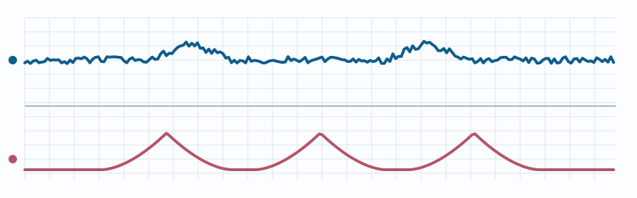
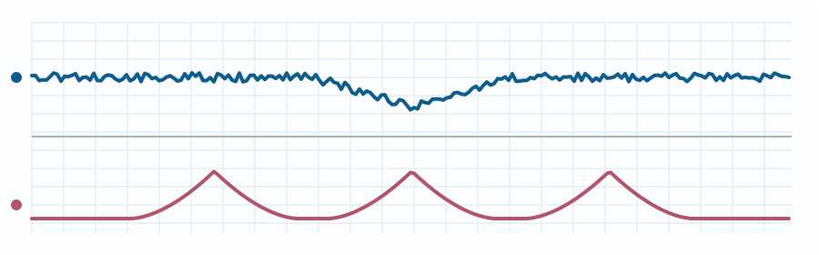
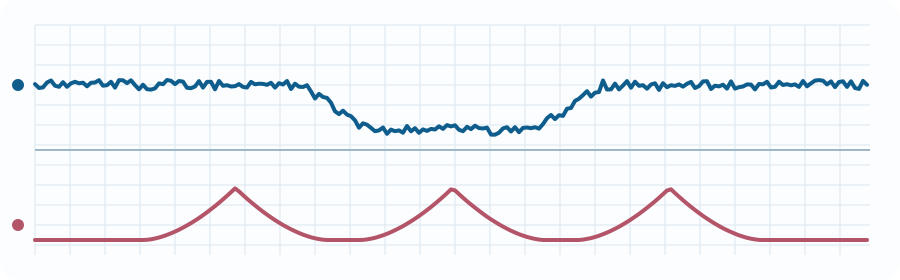
Gradual decreases that mirror contractions. They are often associated with fetal head compression and usually do not require intervention when the rest of the tracing is reassuring.
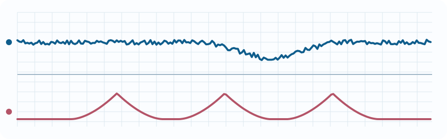
Gradual decreases shifted after the contraction. Recurrent late decelerations can reflect impaired uteroplacental oxygen transfer from maternal hypotension, tachysystole, placental disease, hypoxemia, or other causes.
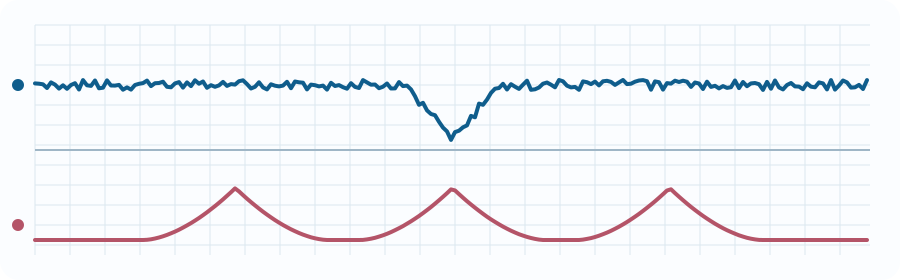
Abrupt decreases with variable timing, depth, and shape. They commonly reflect umbilical cord compression. Recurrent deep or prolonged variables, slow return to baseline, reduced variability, or bradycardia increase concern.
A decrease lasting at least 2 minutes but less than 10 minutes. Causes include maternal hypotension, cord prolapse, uterine rupture, placental abruption, tachysystole, rapid descent, or medication effect. Ten minutes or longer is a baseline change.
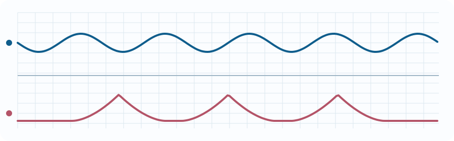
Normal uterine activity is five or fewer contractions in 10 minutes averaged over 30 minutes. Tachysystole is more than five in 10 minutes averaged over 30 minutes. Describe whether decelerations are present. Excessive frequency, prolonged contractions, or inadequate resting tone can reduce uterine perfusion between contractions.
Tachysystole can occur without medication and still requires fetal and maternal evaluation.
Stop or reduce stimulation as authorized when excessive activity or fetal deterioration occurs.
Correct position and perfusion; review epidural, medications, bleeding, and vena-caval compression.
Positioning may help, but persistent patterns require rapid obstetric escalation.
All patterns not Category I or III. Requires continued evaluation, correction of reversible causes, communication, and trend assessment.
Confirm fetal signal and maternal pulse; inspect equipment and transducer position.
Left lateral positioning or uterine displacement when appropriate; relieve cord compression risk.
Correct maternal hypotension, hemorrhage, dehydration, or medication effects according to protocol.
Stop or reduce oxytocin as authorized and address tachysystole.
Support airway and give oxygen for maternal hypoxemia or another clinical indication—not reflexively when saturation is normal.
Notify obstetric leadership, reconsider destination, and prepare for expedited delivery capability.
The 30-week patient has a baseline of 145/min, moderate variability, and no decelerations. This is reassuring for the moment.
After supine loading, maternal blood pressure falls and late decelerations appear. The team confirms the fetal signal, applies left tilt, and treats the maternal perfusion problem.
Blood pressure improves but the tracing retains minimal variability with recurrent late decelerations. The team stops any uterotonic stimulation, reassesses oxygenation and bleeding, contacts the receiving obstetric team, and identifies the nearest surgical-capable destination.
The tertiary center is 35 minutes away and a capable obstetric center is 8 minutes away. Persistent fetal deterioration despite maternal correction supports diversion discussion rather than blind adherence to the original plan.
Immediate rationales are shown in study mode, and your score is stored locally.