IBSC domain: AssessmentsEstimated study time: 110–150 minutesDifficulty: Intermediate–AdvancedClinical review: July 2026
Educational use onlyThis chapter supports certification preparation and transport-clinician reasoning. Follow local protocols, scope of practice, obstetric consultation, and the established transfer plan.
Learning objectives
After completing this chapter, you should be able to distinguish contractions from established labor, describe the stages and phases of labor, assess contraction patterns, explain cervical dilation, effacement, station, presentation, and position, recognize membrane rupture and imminent delivery, identify high-risk presentations, and determine when transport should continue, pause, divert, or convert to delivery preparation.
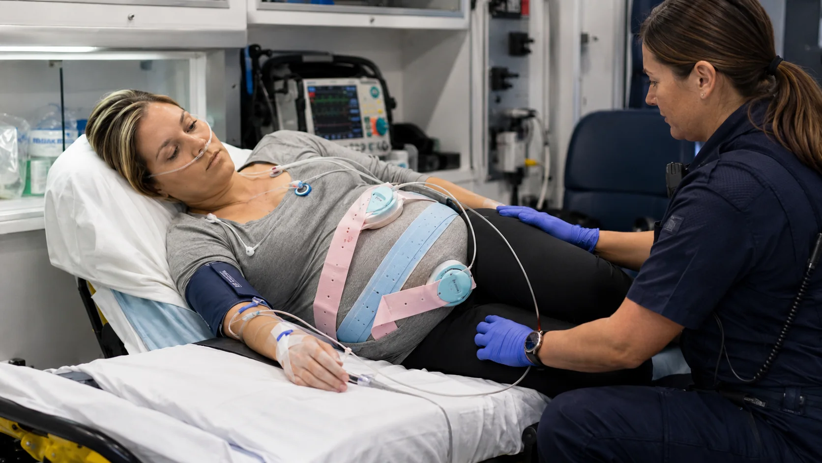
Labor assessment in motion. The transport clinician must integrate contractions, maternal symptoms, membrane status, fetal status, gestational age, and distance to delivery capability. AI-generated clinical training image.
The five labor questions
1
Is this established labor? Contractions plus cervical change—not pain alone.
2
How advanced is it? Stage, dilation, effacement, station, and signs of descent.
3
Is delivery uncomplicated? Presentation, membranes, bleeding, prior uterine surgery, multiples.
4
Is the fetus tolerating labor? Baseline, variability, decelerations, and maternal physiology.
5
Does the destination still fit? Continue, divert, prepare to deliver, or request resources.
Opening transport scenario
A 32-year-old G4P3 at 38 5/7 weeks is being transferred for labor because the sending facility has no obstetric service. Contractions were every 6 minutes at departure. Fifteen minutes later they occur every 2 minutes and last 70 seconds. She reports rectal pressure and says she cannot stop pushing. The perineum bulges during contractions. The closest obstetric-capable hospital is 18 minutes away. This is no longer a routine interfacility ride; the labor assessment now determines whether the safest plan is continued transport or immediate delivery preparation.
Mother–fetus connectionLabor intermittently reduces uterine blood flow during contractions. Excessive uterine activity, maternal hypotension, hypoxemia, fever, or placental disease can reduce fetal reserve and change the urgency of delivery.
1. What counts as labor?
Established labor is uterine contractions that result in cervical dilation or effacement. Contractions can be painful, regular, or frequent without producing cervical change. Membranes can rupture before labor. Conversely, labor may be established before membranes rupture.
More consistent with established labor
Progressive cervical dilation or effacement
Contractions becoming stronger, longer, or closer
Progressive descent and increasing pelvic pressure
Bloody show with other labor findings
Increasing inability to speak or relax through contractions
May be nonlabor uterine activity
Irregular contractions without cervical change
Tightening that decreases with rest, hydration, or position change
No progressive descent or pressure
Stable maternal and fetal findings
No membrane rupture or concerning bleeding
Transport clinicians should not promise that a patient is or is not in labor without the appropriate assessment. When cervical findings are unavailable, document the observable pattern and communicate uncertainty.
2. Stages and phases of labor
1
First stage
Onset of labor to complete cervical dilation. The latent phase precedes active labor; current ACOG guidance uses 6 cm as the start of the active phase.
2
Second stage
Complete dilation to birth. Includes passive descent and active maternal pushing. Crowning and involuntary bearing down suggest imminent birth.
3
Third stage
Birth of the newborn to delivery of the placenta. Monitor bleeding and uterine tone; never apply forceful cord traction.
4
Immediate postpartum
Early recovery after placental delivery. Prioritize uterine tone, bleeding, maternal perfusion, and newborn transition.
Transport decision pointA patient does not need to be fully dilated to deteriorate or require diversion. Advanced labor, abnormal presentation, hemorrhage, prior uterine surgery, fetal deterioration, or an inadequate destination can change the plan at any stage.
3. Contraction assessment
Describe uterine activity using four components:
F
Frequency
Beginning of one contraction to beginning of the next.
D
Duration
Beginning to end of the same contraction.
I
Intensity
Patient report, palpation, or internal measurement when available.
R
Resting tone
Whether the uterus softens adequately between contractions.
Uterine tachysystole is more than five contractions in ten minutes, averaged over thirty minutes. Always report whether fetal heart-rate decelerations accompany the pattern. Tachysystole can occur spontaneously or with oxytocin, prostaglandins, or other stimulation.
Clinical trap“Contractions every two minutes” is not enough. A complete report adds duration, intensity, resting tone, oxytocin or other uterotonic exposure, and the fetal response.
4. Dilation, effacement, station, and descent
Cervical dilation
0–10 cm
Opening of the cervix. Complete dilation is 10 cm.
Effacement
0–100%
Thinning and shortening of the cervix.
Station
−5 to +5
Relationship of the presenting part to the ischial spines; 0 station is level with the spines.
Descent
Trend
Progress of the presenting part through the pelvis. Rapid change can signal imminent birth.
A digital cervical examination is not a routine transport procedure. Perform only when trained, authorized, clinically necessary, and safe. Avoid digital examination in unexplained third-trimester bleeding until placenta previa has been excluded. Repeated examinations increase infection risk after membrane rupture.
5. Lie, presentation, and position
Cephalic
Head first. Vertex is the usual favorable presentation.
Breech
Buttocks or feet first. Higher risk for cord prolapse, head entrapment, and neonatal compromise.
Transverse/oblique
Shoulder or side presents. Vaginal birth is generally not possible without change in lie.
Compound/limb
An extremity accompanies or precedes the presenting part. Do not pull on a visible limb.
Fetal position describes the orientation of a reference point—usually the occiput in a vertex fetus—within the maternal pelvis. Exact position may be documented by the obstetric team, but transport clinicians should at minimum know presentation, whether the presenting part is engaged or descending, and whether a cord or extremity is visible.
6. Membrane status and fluid assessment
Document the time of rupture, whether rupture was spontaneous or artificial, and the amount, color, odor, and ongoing leakage of fluid. Clear fluid may be normal. Green or brown fluid suggests meconium. Foul odor with maternal fever or uterine tenderness raises concern for infection. Blood-stained fluid requires evaluation for bleeding causes.
Cord prolapse
Visible or palpable cord, sudden fetal bradycardia, or recurrent variable decelerations after rupture.
Meconium
Signals fetal stool passage and need for neonatal readiness; it does not by itself prove acidemia.
Infection
Fever, foul fluid, uterine tenderness, maternal or fetal tachycardia.
Preterm rupture
Requires gestational-age-specific obstetric and neonatal capability.
7. Signs of imminent delivery
Crowning or visible presenting part
Involuntary bearing down or inability to stop pushing
Strong rectal pressure or urge to have a bowel movement
Perineal bulging or gaping
Rapidly increasing contraction frequency and intensity
Progressive descent or a presenting part at a low positive station
A multiparous patient reporting that birth feels immediate
Prepare for delivery before the final sign appears. Establish a warm environment, newborn resuscitation readiness, maternal hemorrhage supplies, suction, oxygen, clamps, towels, and additional help.
8. High-risk labor findings
Prior uterine surgery
Sudden constant pain, fetal deterioration, bleeding, shock, or loss of station may indicate rupture.
Breech or transverse lie
Requires immediate obstetric capability and careful cord assessment.
Multiple gestation
Identify each fetus, anticipate malpresentation and more than one newborn, and prepare for hemorrhage.
Preterm labor
Gestational age changes medication, neonatal, destination, and delivery decisions.
Hemorrhage
Painful bleeding, uterine tenderness, or shock may indicate abruption or rupture.
Epidural complications
Hypotension can reduce uteroplacental perfusion and trigger fetal deterioration.
9. Continue, divert, or prepare to deliver
Continue with frequent reassessment
Stable maternal and fetal status, no imminent birth, appropriate destination and resources, adequate travel margin.
Pause before movement
Unsecured equipment, incomplete assessment, active resuscitation, uncontrolled hemorrhage, uncertain presentation, or absent delivery readiness.
Prepare to deliver
Crowning, involuntary pushing, visible presenting part, rapidly advancing second stage, or unsafe remaining travel time.
Divert/escalate
Fetal deterioration, maternal instability, abnormal presentation, uterine rupture concern, cord prolapse, or need for immediate surgical/neonatal capability.
10. Evolving case study
Phase 1: Initial transfer
The G4P3 patient has contractions every 6 minutes, reassuring fetal status, intact membranes, and no urge to push. The original obstetric destination is appropriate.
Phase 2: Accelerating labor
Contractions become every 2 minutes and last 70 seconds. The uterus softens between contractions, but the patient reports strong rectal pressure. The team reassesses the perineum, confirms fetal status, and notifies the receiving center.
Phase 3: Imminent birth
Crowning and involuntary pushing develop. The team stops in the safest feasible location, warms the compartment, opens delivery and neonatal equipment, assigns maternal and newborn roles, and prepares for birth.
Phase 4: Postbirth transport
After birth, the newborn is dried, warmed, assessed, and supported. The mother is monitored for placental delivery, uterine tone, and hemorrhage. The team avoids forceful cord traction and continues to the appropriate receiving facility.
11. High-yield chapter summary
Labor is contractions with cervical change.
Active labor generally begins at 6 cm.
Report contraction frequency, duration, intensity, and resting tone.
Tachysystole is more than five contractions in ten minutes averaged over thirty minutes.
Complete dilation is 10 cm; 0 station is at the ischial spines.
Crowning, involuntary pushing, and perineal bulging indicate imminent birth.
Assess membrane status, fluid, and cord prolapse risk.
Avoid digital examination in unexplained late-pregnancy bleeding.
Prior uterine surgery changes the differential for sudden pain and fetal deterioration.
Abnormal presentation, preterm labor, multiples, and hemorrhage change destination needs.
The transport plan must change when labor advances faster than travel time.
Never apply forceful traction to deliver the placenta.
References
International Board of Specialty Certification. Maternal Fetal Transport Microcredential Candidate Handbook. Updated April 2026.
American College of Obstetricians and Gynecologists. First and Second Stage Labor Management. Clinical Practice Guideline No. 8. 2024.
American College of Obstetricians and Gynecologists. reVITALize: Obstetrics Data Definitions.
American College of Obstetricians and Gynecologists. Cervical Ripening in Pregnancy. Clinical Practice Guideline No. 9. 2025.
Macones GA, et al. The 2008 NICHD workshop report on electronic fetal monitoring. Obstet Gynecol. 2008;112:661–666.