Lung aeration
Air enters the lungs and creates functional residual capacity.
After completing this chapter, you should be able to:
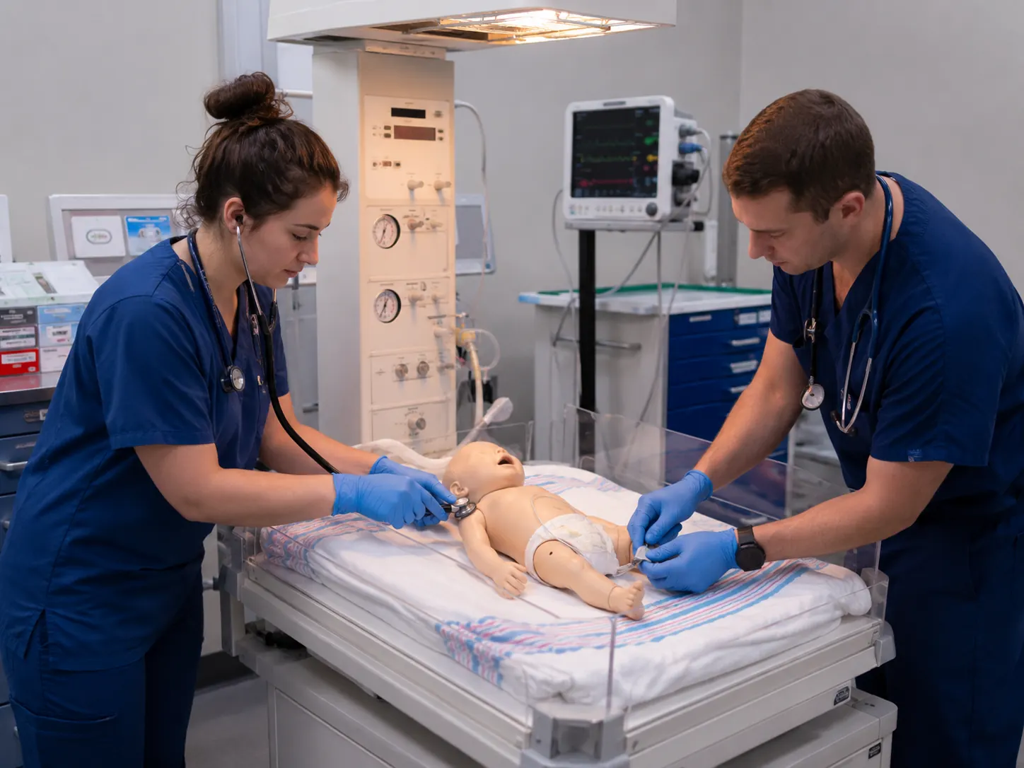
Term gestation?
Prematurity predicts reduced respiratory and thermal reserve.
Good tone?
Poor tone may reflect hypoxia, prematurity, medication exposure, infection, or neurologic compromise.
Breathing or crying?
Apnea, gasping, or ineffective effort requires rapid airway and ventilation support.
A 34-week newborn is delivered shortly before the transport team reaches a rural emergency department. The newborn is limp, has irregular gasping respirations, and a heart rate of 88/min. The room is cool, the infant is wet, and no blended oxygen source is connected. This is not primarily a “cardiac” problem. The most likely immediate failure is inadequate lung inflation and gas exchange during the transition from placental to pulmonary support.
Before birth, the placenta performs gas exchange, the lungs are fluid filled, pulmonary vascular resistance is high, and the fetal shunts direct blood away from the pulmonary circulation. At birth, the newborn must establish ventilation, replace lung fluid with air, increase pulmonary blood flow, redirect cardiac output, maintain body temperature, and begin independent glucose regulation—often within minutes.[1,2]
Air enters the lungs and creates functional residual capacity.
Oxygenation and lung expansion reduce pulmonary vascular resistance.
More right-ventricular output reaches the lungs.
Pulmonary venous return promotes functional closure of the foramen ovale.
Placental support is replaced by pulmonary gas exchange.
The fetal lungs contain liquid. During labor and after birth, fluid is cleared through epithelial sodium transport, thoracic recoil, lymphatics, and pulmonary blood vessels. The first effective breaths require enough pressure to move fluid, open alveoli, and establish a stable volume of air remaining after exhalation—functional residual capacity (FRC).
Two events drive the cardiovascular transition: lung aeration lowers pulmonary vascular resistance, and umbilical cord clamping removes the low-resistance placental circulation and raises systemic vascular resistance. These changes alter pressure relationships across the fetal shunts.
Increased pulmonary venous return raises left-atrial pressure. The septal flap functionally closes when left pressure exceeds right pressure.
Rising oxygen tension and falling prostaglandin influence promote constriction. Persistent hypoxemia or acidosis can delay closure or reopen shunting.
Umbilical flow stops after cord clamping. The venous bypass of the liver is no longer required and closes over time.
Current neonatal resuscitation guidance emphasizes preparation before birth, immediate assessment, thermal protection, and timely ventilation. The newborn’s heart rate is the most useful objective measure of response. Auscultation is used initially; electrocardiography provides rapid and accurate continuous heart-rate assessment during active resuscitation, while pulse oximetry is required to guide oxygen therapy.[1]
≥100/min: generally reassuring when breathing is effective.
<100/min: evaluate ventilation and provide positive-pressure ventilation when indicated.
<60/min: advanced escalation after adequate ventilation.
Assess apnea, gasping, crying, rate, retractions, grunting, symmetry, and air entry. Gasping is not effective breathing.
Flexion and spontaneous movement are reassuring. Hypotonia may reflect prematurity, hypoxia, maternal medication, infection, or neurologic injury.
Central cyanosis, pallor, delayed capillary refill, weak pulses, and poor response to ventilation require rapid reassessment.
For a newborn who is not transitioning normally, initial actions are to warm and maintain normal temperature, dry when appropriate, position the airway, stimulate, and clear the airway only when needed. A newborn who remains apneic or gasping, or whose heart rate remains below 100/min after initial steps, should receive assisted ventilation within the first 60 seconds after birth.[1]
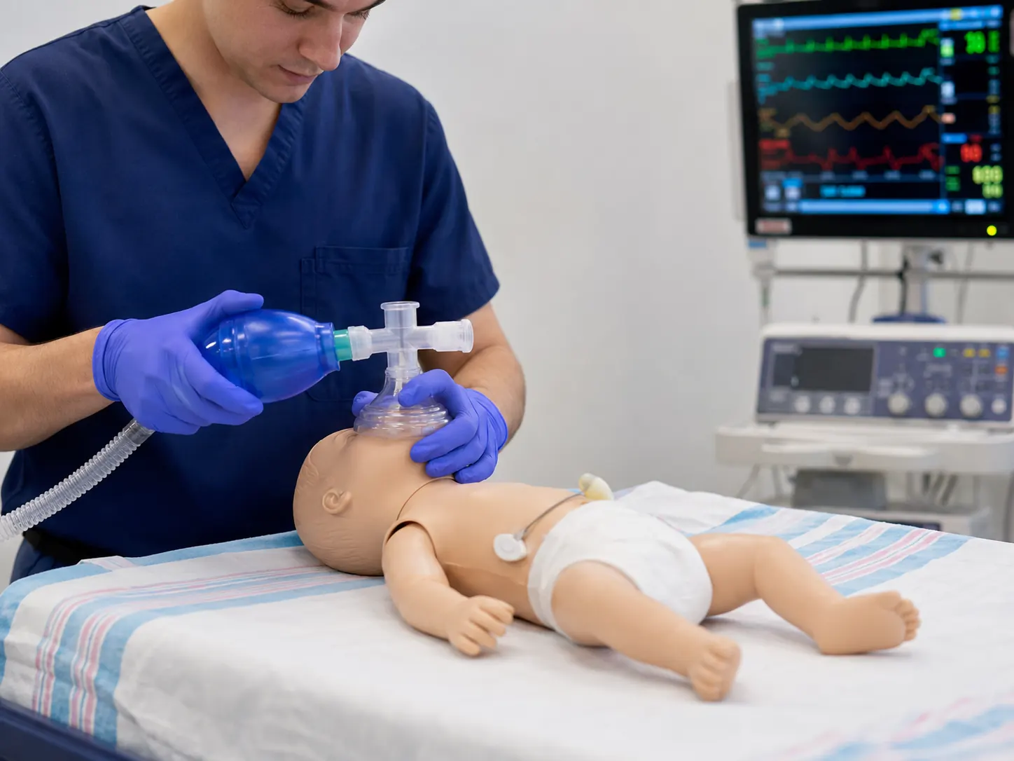
Ventilation may begin with 21% oxygen in term and late-preterm newborns. Current guidance recommends 21%–30% oxygen for infants approximately 32–35 weeks and individualized higher starting concentrations for very preterm infants, with adjustment using preductal pulse oximetry and target saturations.[1] Follow your local NRP implementation and equipment capabilities.
| Time after birth | Target SpO₂ |
|---|---|
| 1 minute | 60%–65% |
| 2 minutes | 65%–70% |
| 3 minutes | 70%–75% |
| 4 minutes | 75%–80% |
| 5 minutes | 80%–85% |
| 10 minutes | 85%–95% |
Place the pulse-oximetry sensor on the right hand or wrist to measure preductal saturation. Normal transition is gradual; do not force immediate adult saturation values with unnecessary oxygen.[1]
If the heart rate does not rise, correct ventilation before advancing. Consider mask repositioning, airway repositioning, suction when obstruction is suspected, opening the mouth, increasing pressure judiciously, and placement of an alternative airway. Chest compressions are indicated when the heart rate remains below 60/min despite at least 30 seconds of ventilation that moves the chest, preferably through an endotracheal tube or laryngeal mask. Compressions are coordinated at a 3:1 ratio with ventilation. Vascular access and epinephrine are considered when the heart rate remains below 60/min despite effective ventilation and compressions.[1]
Newborns lose heat rapidly because of a large surface-area-to-mass ratio, wet skin, limited insulation, and limited ability to generate heat. Cold stress increases oxygen and glucose consumption and can worsen pulmonary vasoconstriction, hypoglycemia, and metabolic acidosis. The goal is normothermia—not overheating. WHO defines hypothermia as a temperature below 36.5°C; sick and preterm newborns require continuous thermal support and temperature monitoring.[3]
Heat is lost as amniotic fluid evaporates. Dry promptly unless using a preterm plastic-wrap strategy.
Air currents remove heat. Close doors, reduce drafts, and warm the patient compartment.
Contact with cold surfaces causes heat transfer. Prewarm blankets, mattress, and equipment.
Heat radiates to nearby cold surfaces without direct contact. Use a warmer or transport isolette.
After the cord is clamped, continuous maternal glucose delivery stops. The newborn must mobilize glycogen, use fat stores, and establish feeding. Prematurity, hypothermia, sepsis, maternal diabetes, growth restriction, respiratory distress, and prolonged resuscitation increase the risk of hypoglycemia. Postresuscitation monitoring should include blood glucose when indicated, but treatment thresholds and infusion strategies must follow neonatal protocols and medical direction.
The Apgar score describes the newborn’s condition and response to intervention at defined time points. It is assigned at 1 and 5 minutes; if the 5-minute score is below 7, scoring is typically repeated every 5 minutes through 20 minutes. The score must not delay resuscitation and should not be used alone to predict an individual infant’s neurologic outcome.[4]
| Component | 0 | 1 | 2 |
|---|---|---|---|
| Appearance | Blue or pale | Body pink; extremities blue | Completely pink |
| Pulse | Absent | <100/min | ≥100/min |
| Grimace | No response | Grimace | Cough, sneeze, or vigorous response |
| Activity | Limp | Some flexion | Active motion |
| Respiration | Absent | Slow or irregular | Good cry |
Maternal medications may alter neonatal transition. The transport clinician should obtain the medication, dose, route, timing, and maternal renal function whenever relevant.
Anticipate hypotonia, weak respiratory effort, and depressed reflexes, particularly with high maternal levels or impaired renal clearance. Prioritize ventilation and supportive care.
Respiratory depression may occur. Effective ventilation remains the immediate priority. Antagonist use requires neonatal-specific consultation and awareness of maternal dependence.
May contribute to reduced tone and respiratory effort. Prepare for airway support and prolonged observation.
Monitor heart rate and glucose closely according to receiving-facility guidance.
Newborns who require more than routine initial steps are at risk for recurrent apnea, hypoxemia, hypoglycemia, hypothermia, abnormal tone, seizures, and hemodynamic instability. Reassessment must continue during every movement and equipment change.[1]
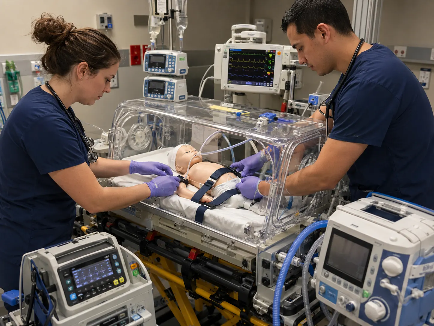
The 34-week newborn remains apneic with a heart rate of 88/min after warming, positioning, drying, and stimulation. The team begins positive-pressure ventilation with an appropriately sized mask and blended oxygen. There is minimal chest movement and the heart rate remains 86/min.
Interpretation: Ventilation is not yet effective. Reposition the airway, improve the mask seal, open the mouth, assess for obstruction, adjust pressure to achieve lung inflation, and consider an alternative airway if the heart rate does not rise.
After corrective steps, the chest moves gently and the heart rate rises to 132/min. Spontaneous respirations begin, but the infant has grunting and retractions. Preductal SpO₂ is rising through the expected transitional range.
Interpretation: The rise in heart rate confirms effective ventilation. A spontaneously breathing preterm newborn with ongoing work of breathing may benefit from continuous positive airway pressure according to protocol and available expertise.
Twenty minutes later, the infant’s axillary temperature is 35.9°C and glucose is trending down. The transport isolette has not been preheated.
Decision: Correct the thermal environment before departure, initiate glucose management according to neonatal orders, and reassess respiratory support. Hypothermia increases oxygen and glucose consumption and can destabilize an infant who initially appeared improved.
Immediate rationales are shown in study mode, and your final score is stored locally.